
HIP LABRUM TEAR, CARTILAGE TEAR
Diagnostic hip arthroscopy showing cartilage tear and labral tear
WHAT IS THE HIP LABRUM?
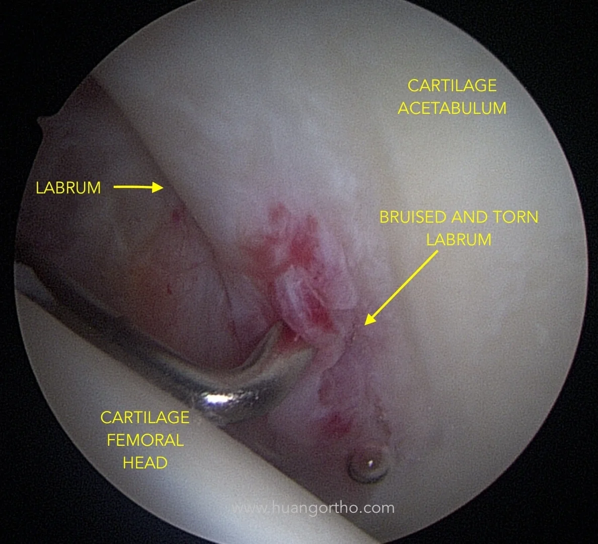
Arthroscopic probe in area of hip labrum that is torn and bruised. Adjacent labrum is normal as is the cartilage on the acetabulum (socket) and femoral head (ball).
The labrum is a structure on the acetabular (socket) rim. Imagine the socket as a margarita glass. The labrum would be the salt around the rim of the glass, but instead of the grains of salt, the labrum is a ring of fibrous tissue. The function of the labrum is not completely understood, but it does serve to protect the cartilage next to it and seal the joint. It has many nerve endings in it so if it becomes damaged, it can be a source of pain. In the setting of femoroacetabular impingement (FAI), the hip labrum see abnormal contact from the bony collision which can be a cause of labral tears.
WHAT IS HIP CARTILAGE?
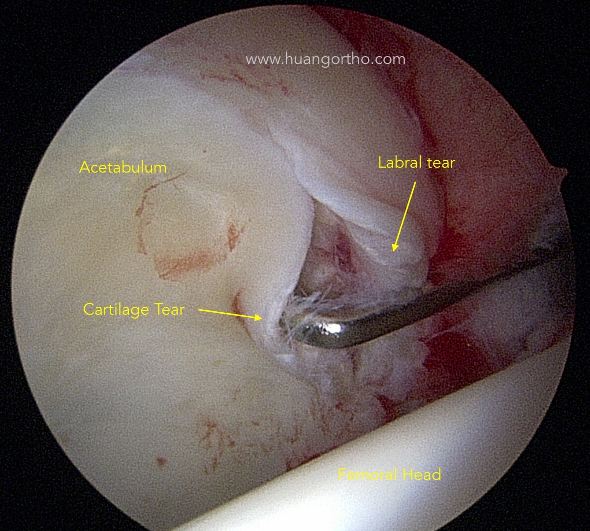
Arthroscopic photo demonstrating hip cartilage tear and labral tear along with adjacent normal cartilage of acetabulum (socket) and femoral head (ball).
Cartilage is the structure in our joints that gives us a smooth, low friction surface to walk on. It is found on both the femoral head and the acetabulum. The cartilage layers in our hips can tear with continued trauma from femoroacetabular impingement (FAI). Arthritis can be defined as the loss of cartilage in our joints. So as FAI continues, then the cartilage damage can progress effectively increasing the arthritis in the hip.

Hip cartilage tear seen adjacent to large hip labral tear.

Closer view of large hip cartilage tear. Easily displaceable upon arthroscopic probing.
SYMPTOMS of labral tear and/or cartilage tear may include:
Groin pain associated with hip activity
Front, side, buttock pain location
Pain can be dull, achy, or sharp and knife-like
Mechanical symptoms may include locking, catching, giving way
Pain often radiates to front or inner thigh but does not typically go below the knee
Pain is worse with sitting, squatting, walking up or downhill
Range of motion may decrease

MRI Arthrogram showing complete labral tear
DIAGNOSIS is made with a combination of:
Patient history
Physical exam
X-rays
MRI
TREATMENT options include conservative (non-surgical) and surgical:
Conservative treatment options refer to management of the problem without surgery. Nonsurgical management of FAI will probably not change the underlying abnormal biomechanics of the hip causing the FAI but may offer pain relief and improved mobility.
Rest
Activity modification and limitations
Anti-inflammatory medication
Physical therapy
Injection of steroid
Injection of biologics: platelet rich plasma or stem cell
Surgical treatment:
Hip arthroscopy (minimally invasive surgery) is reasonable to consider if conservative treatments have failed. Surgery is designed to repair the labrum back to the acetabulum and address any cartilage damage. At the same time, any bony impingement is removed to help prevent recurrent tear. Occasionally, the labrum cannot be fixed or has failed prior surgical repair. In this situation, labral reconstruction (making a new labrum with other tissue) can be considered.

Arthroscopic probe testing hip labral repair
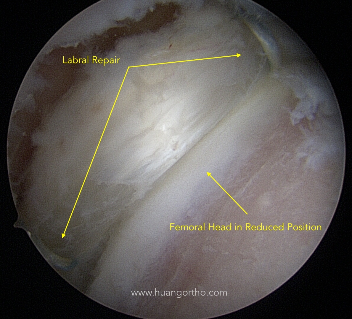
Femoral head in reduced position and labral repair in anatomic position recreating the "seal" of the hip joint.